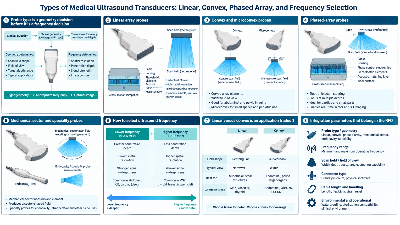
Probe type is a geometry decision before it is a frequency decision
A medical ultrasound transducer converts electrical excitation into acoustic pulses and returning echoes into electrical signals. The probe type determines how elements are arranged, how the beam is steered and what field of view reaches the system. Frequency then trades penetration against spatial resolution within that geometry.
For an OEM project, “linear or convex” is not enough. Define clinical application, target depth, access window, footprint, channel count, connector, beamforming architecture, acoustic output limits, cleaning method and regulatory pathway. A transducer is one component of a medical device and does not establish diagnostic performance by itself.
Linear array probes
A linear array has elements arranged along a straight aperture and normally produces a rectangular image. Its broad near-field contact and parallel scan lines suit superficial structures, vascular access, musculoskeletal imaging and other applications requiring fine near-surface detail.
Linear probes often operate at higher frequencies than general abdominal probes. That improves resolution but reduces penetration. The footprint may be too large for narrow acoustic windows, and element pitch must be controlled to manage grating lobes when steering.
Convex and microconvex probes
A convex array places elements along a curved aperture, producing a sector-like image that widens with depth. It offers broad coverage from a moderate contact area and is commonly associated with abdominal, obstetric and general imaging. The BBMU-ABD abdominal B-mode array probe illustrates an OEM component boundary for this class.
A microconvex probe uses a smaller curved footprint for limited access, pediatric or veterinary configurations. The reduced aperture changes beamwidth and resolution, so it is not simply a smaller housing around the same acoustic design.
Phased-array probes
A phased array uses a compact aperture and electronic timing delays to steer and focus a sector beam. Its small footprint helps access between ribs and other narrow windows. The system requires tightly controlled element behavior, channel timing and beamforming.
Element count alone does not predict image quality. Pitch, aperture, bandwidth, matching layers, backing, crosstalk, cable and front-end electronics all affect the pulse and received echo. Confirm the transducer and beamformer as a matched architecture.
Mechanical sector and specialty probes
A mechanical sector probe moves or rotates one or more elements to sweep the beam. This can reduce electronic channel count while producing a sector image, but adds motors, seals, bearings, motion control and service-life considerations. The BBMU-BS mechanical sector-scan transducer is an example of this distinct integration route.
Endocavity, transesophageal, intraoperative, catheter and other specialty probes are shaped by access, sterilization or disinfection, patient-contact materials and cable mechanics. Their constraints often dominate the acoustic selection.
How to select ultrasound frequency
Higher frequency shortens wavelength and can improve axial resolution, but tissue attenuation increases and useful depth falls. Lower frequency penetrates farther but cannot resolve the same small features. The correct operating band must cover the target depth while preserving acceptable resolution and signal-to-noise ratio.
Do not choose from center frequency alone. Specify fractional bandwidth, pulse length, sensitivity, impedance, element uniformity and operating modes. Harmonic imaging, Doppler and pulsed modes may impose different bandwidth and thermal requirements.
Linear versus convex is an application tradeoff
Choose linear when the target is shallow and a rectangular near-field view is useful. Choose convex when wider deep coverage and a curved contact surface better suit the anatomy. If access is narrow and electronic steering is needed, phased array may be the better architecture. The choice follows the acoustic window and required view, not a universal ranking.
Integration parameters that belong in the RFQ
- Application, target depth, field of view and desired footprint.
- Center frequency, bandwidth, element count, pitch and aperture.
- System channels, excitation, receive impedance and connector pinout.
- Matching layer, lens, backing, cable length and strain relief.
- Housing dimensions, patient-contact materials and biocompatibility needs.
- Cleaning, disinfection, sterilization and ingress requirements.
- Acoustic, electrical, thermal and lifetime acceptance tests.
Prototype evaluation should use the intended front end, beamformer, cable, acoustic load and operating modes. A good pulse-echo result on a generic bench does not complete medical-device verification.
The FDA overview of ultrasound imaging provides a regulatory and safety starting point for medical-device development, including the principle of using the lowest exposure needed to obtain the required information. Translate the application into a controlled specification with the custom transducer RFQ guide, establish component evidence with the testing guide, and coordinate the electrical interface with the circuit-matching guide.
FAQ
Which probe gives the highest resolution?
There is no universal answer. Higher frequency generally improves resolution, but usable resolution depends on depth, aperture, focusing, bandwidth, system processing and the acoustic path.
What is the main difference between linear and convex probes?
Linear probes usually provide a rectangular view and strong near-field access; convex probes provide a widening field for broader coverage at depth.
Is a phased array the same as a convex array?
No. Both can produce sector-shaped images, but the element arrangement, footprint and steering method differ.
Can a probe be connected to any ultrasound system?
No. Electrical architecture, channel count, pinout, identification, beamforming, software and safety requirements must all be compatible.